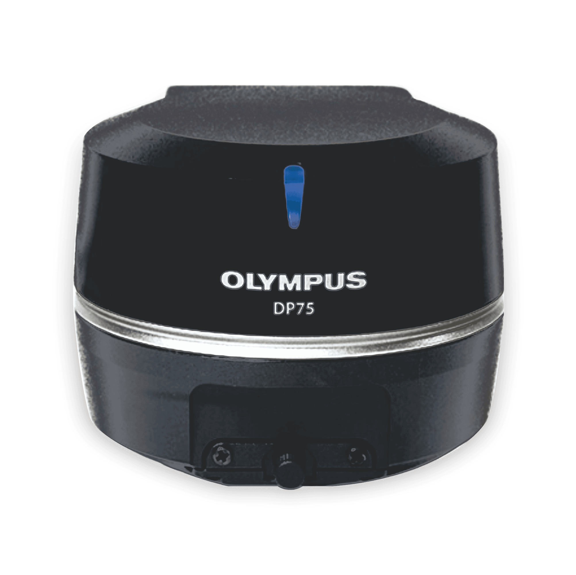
Microscope Digital Camera DP75
One Camera. Multiple Applications
The DP75 digital microscope camera is a high-performance, multi-application imaging tool that makes it easy to capture high-resolution brightfield or fluorescence images using a single camera. It simplifies your microscopy imaging, so you can focus more on your work.


Sharper Microscopy Images, Clearer Insights
The DP75 camera makes it easier than ever to capture sharp, low-noise images. Its exceptional color reproduction makes your images as vivid as looking through the microscope oculars.
Rat Pancreas. Stain: DAPI AF555 Cy5
Colon. Stain: HE
Ptk2. Stain: Cy7
Infrared Fluorescence Imaging
The DP75 camera supports multiple staining combinations and wavelengths up to 1000 nm with a switchable infrared (IR) cut filter. This setup enables you to—for example—check your sample conditions with your standard widefield microscope before spending time on a confocal microscope to finalize your imaging.
Qualitative Analysis Capabilities
Dive deeper with the camera’s advanced analysis capabilities.
- Linear mode enables relative intensity evaluation on a fluorescence image so that you can quantitatively compare the intensity of fluorescence expressions.
- Easily overlay fluorescence and brightfield images with pixel precision since you are using the same sensor for brightfield and fluorescence. Precisely identify the location of fluorescent expression, helping you focus on the relevant morphology and localization of your specimen.
Image provided by Dr. Hiroyuki Nakajima, National Cerebral and Cardiovascular Center
Smart Observation Detection
The AI-based scene detection feature automatically recognizes five observation methods (brightfield, fluorescence, phase contrast, differential interference contrast, and polarization), enabling anyone to obtain high-quality images with minimal training.
The DP75 is not for clinical diagnostic use.
Fast Frame Rate
When imaging live specimens, a fast frame rate is important for efficiency and capturing the dynamics of your samples. With a fast frame rate of 22 frames per second (fps) at over 4K resolution and 60 fps at full HD resolution, the camera provides smooth, fast, live images for easy framing and comfortable live observation.
Top right: Without pixel shift / Bottom right: With pixel shift
High-Resolution, Wide Field of View Imaging
The camera’s wide field of view capabilities enable you to find your target areas quickly, making your research more efficient. The DP75 camera also enables you to capture high-resolution images even at low magnification with a maximum resolution of 8192 × 6000 through pixel shifting modes.
Multiple Image Alignment (MIA) Capabilities
The instant multiple image alignment (MIA) function simplifies the creation of wide-area images by moving the XY stage manually without any motorized setup, and the integrated position navigator helps ensure that you always know your position on the sample during brightfield and fluorescence imaging.
Flexible Upgrades
Since the DP75 camera uses USB 3.1 Gen2, it is compatible with most PCs for a simple, effective upgrade to your current system.
The DP75 is not for clinical diagnostic use.
| Camera type |
|
|---|---|
| Image Sensor | 1.1 inch 12.37 megapixel color CMOS image sensor, global shutter |
| Resolution (max.) | 8192 (W) × 6000 (H) pixels |
| Pixel Size | 3.45 x 3.45 µm |
| Effective image resolution |
|
| Sensitivity |
|
| A/D Converter | 12 bits |
| Binning | 2 × 2 |
| Metering modes |
|
| White balance | Auto / One-touch / Manual / Area designation |
| Black balance | Auto / One-touch / Manual / Area designation |
| Exposure Times | 28 μs–120 s |
| Live Frame Rates |
|
| Still image transfer time |
|
| Monochrome mode | Available (Standard / Custom) |
| Color space | sRGB, AdobeRGB 2 |
| Linear mode | Available |
| IR-cut filter | Switchable
|
| Manual panoramic imaging (Instant MIA) | Available (Supporting fluorescence as well as brightfield) 3 4 |
| Auto scene recognition mode | Available by AI algorithm (Supporting scene : brightfield, fluorescence, phase contrast, differential interference contrast, and polarization) 4 |
| Position navigator | Available 4 |
| Control Software | cellSens Entry / Standard / Dimension Ver 4.2.1 or later DP2-TWAIN Ver 10.5 or later |
| Cooling System | Peltier cooling |
| External Trigger | Available (Input / Output) |
| Data Transfer | USB 3.1 Gen2 (Type-C) |
| Partial Readout | ✓ |
| PC Control |
|
| PC Requirements | CPU: Intel Core i5, Intel Core i7, Intel Xeon, or equivalent RAM: 8 GB or more(recommended 16 GB or more) PC I/F: USB 3.1 Gen2(TypeA) (a dedicated board is unnecessary) 5 |
| Dimensions | Camera interface cable: Approx. 2.7 m AC adaptor: 107 (W) x 47 (D) x 30 (H) mm / Approx .0.3 kg Controller: 384(W)×308(D)×100(H)mm |
- Frame rate may decrease depending on the condition of your PC, monitor resolution, and/or software.
- Monitor designed to meet Adobe RGB is required.
- Manual Process option license is required for cellSens Standard.
- Not available in the combination of cellSens Entry or DP2-TWAIN.
- Operatable with USB3.1 Gen1 (5 Gbps), but framerate is decreased.
